


")
Breast Ultrasound
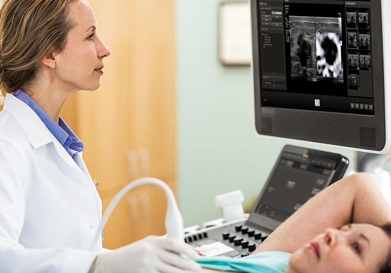
Ultrasound examination of the breasts is an imaging method, which contributes significantly to the diagnosis of breast disorders.
In principle, it works in addition to mammography and helps the specialist to clarify whether a finding of mammography is solid or cystic. Because it is a radiation-free test, breast ultrasound is also used in young girls aged 20-21 years and older who should not be breast-fed because the breasts are sensitive to radiation and their diagnostic value is low due to increased density of glandular tissue.
It also occurs in pregnant women, regardless of the stage of pregnancy, to control the breasts when there is a palpable finding that needs to be investigated.
Ultrasound scanning also helps detect non-palpable lesions, i.e. lesions that are of very small size or in a position that does not allow them to be highlighted by the clinical examination by the doctor.
The ultrasound detects such lesions, but can be used simultaneously for directional a needle biopsy, in which material is taken and a cytological examination is performed to see what is happening. Finally, when there is nipple discharge and especially bloody, ultrasound contributes to not detecting intraductal lesions, i.e. lesions detected in lactiferous porous.
Ultrasound breast should be:
- In dense breasts.
- When suspicious mammogram mass is detected, non-palpable.
- In palpable mass and tissue anarchy.
- When there is a heavy family history.
- Aged under 30 (for screening).