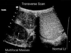
Ultrasound scrotum

It is a non-invasive test used to assess the testicles and epididymis.
It is performed with an ultrasound machine, to which a special head has been fitted that allows the examination of these organs.
Most of today's machines also have Doppler technology, which measures blood flow to the testicles. The examination is performed by a radiologist or urologist who is trained in ultrasound of the urinary system.
There are many reasons why your doctor may recommend that you have the test:
- In the investigation of a palpable mass (such as cancer) or in testicular pain.
- In inflammation of the testicles or epididymis.
- If testicular torsion is suspected (Doppler will show no blood flow).
- In the diagnosis of presence of fluid in the scrotum (hydrocele), blood (hemorrhage) or pus.
- In infertility (such varicose veins).
The procedure that will follow is:
- You will lay down on the examine bed, after removing your clothes from the middle and bottom, in a supine position.
- A clear thick gel will be applied to the examination site. You should be aware that any residue left, does not stain your clothes.
- The doctor will then apply the ultrasound head on the gel and begin to scan the area. You might be asked to take a deep breath and hold it to push it down, increasing the pressure on the abdomen.
- This is done to evaluate the varicose vein and the regression of venous blood to the testicles.
- After the test, the doctor will help to wipe excess gel.


